Anatomy Of Musckes Sndctendons / Images 05. Muscular System | Basic Human Anatomy - This is lesson 1 on the anatomy of the forearm.. The right scapula from the front and back side. Find the best weight lifting exercises that target each muscle or groups of muscles. Tendons are the connective tissues that transmit the mechanical force of muscle contraction to the bones; Tendons vary in size and are somewhat elastic and attach bones to muscles. The quad muscles— which form the meaty mass on the front of your thighs — are among your strongest muscle groups, and play a critical role in athletic activities.
This video also provides you with a. Lying exposed between the protective bones of the superiorly located ribs and the inferiorly located pelvic girdle, the muscles of this region play a critical role in protecting the. Most of the muscles which act on the wrist joint are situated within the forearm, with only the tendon crossing the joint and inserting on the hand. Skeletal muscles are attached to bones by tendons and can be as long as 30 cm, although they are usually 2 to 3 cm in length. There are three layers of gluteal muscles on the posterior hips, just like there are three layers of muscles in the abdominal trunk.
Muscles Of Upper Limb Photograph by Asklepios Medical Atlas from images.fineartamerica.com By connecting our rigid bones to our powerful muscles, tendons allow us to move. The calf muscles (gastrocnemius and soleus), which are connected to the calcaneus via the achilles tendon. On the other hand, the insertion is where a tendon attaches that muscle to the *more* movable bone. The quadriceps muscles provide strength and power with knee extension (straightening). Together, these muscles straighten your knee, stabilize your knee joint, assist in flexing your hip (drawing your knee towards your chest), and help absorb force when you land after jumping or leaping. The foot consists of thirty three bones, twenty six joints and over a hundred muscles, ligaments and tendons. Extensor carpi radialis brevis extensor carpi radialis longus Foot and ankle anatomy is quite complex.
This video also provides you with a.
This video also provides you with a. Related posts of muscles and tendons of the leg muscle anatomy forearm. The quadriceps muscles provide strength and power with knee extension (straightening). These all work together to bear weight, allow movement and provide a stable base for us to stand and move on. Many of the muscles that move the fingers and thumb originate in the forearm. *the origin, insertion, and belly.* a muscle's origin is where a tendon attaches it to the *less* movable bone. The red lines show where the tendons attach the muscles to the bones. When the muscle contracts, the tendons are pulled, and the bone is moved. The hands enable us to perform many of our daily activities such as driving, writing and cooking. The quad muscles— which form the meaty mass on the front of your thighs — are among your strongest muscle groups, and play a critical role in athletic activities. Hochwertiges kletterzubehör für dein outdoor abenteuer! Tendons are the connective tissues that transmit the mechanical force of muscle contraction to the bones; It is comprised of two bones:
It is comprised of two bones: The quadriceps muscles provide strength and power with knee extension (straightening). Ligaments and tendons are fibrous bands of connective tissue that attach to bone. The quad muscles— which form the meaty mass on the front of your thighs — are among your strongest muscle groups, and play a critical role in athletic activities. Human anatomy and physiology lab (bsb 141) module 9:
Muscles Of Right Upper Arm Photograph by Asklepios Medical ... from images.fineartamerica.com Muscles of the hips and thighs. The muscles you probably know the best are your glutes. The posterior tibialis muscle, which supports the arch of the foot and enables the foot to turn. Nerves and blood vessels that supply the bones and muscles of the hip. The quadriceps muscles provide strength and power with knee extension (straightening). The quad muscles— which form the meaty mass on the front of your thighs — are among your strongest muscle groups, and play a critical role in athletic activities. The human hand is made up of the wrist, palm, and fingers and consists of 27 bones, 27 joints, 34 muscles, over 100 ligaments and tendons, and many blood vessels and nerves. The muscular system consists of about 700 muscle organs that are typically attached to bones across a.
Tendons vary in size and are somewhat elastic and attach bones to muscles.
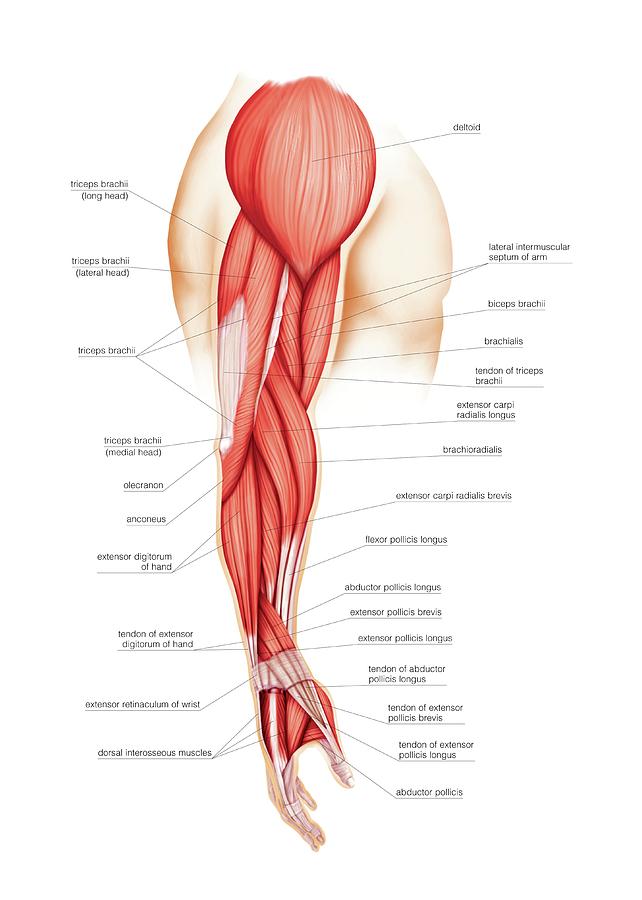
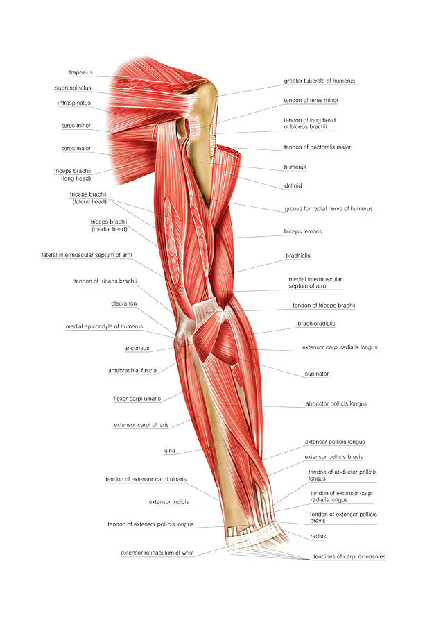
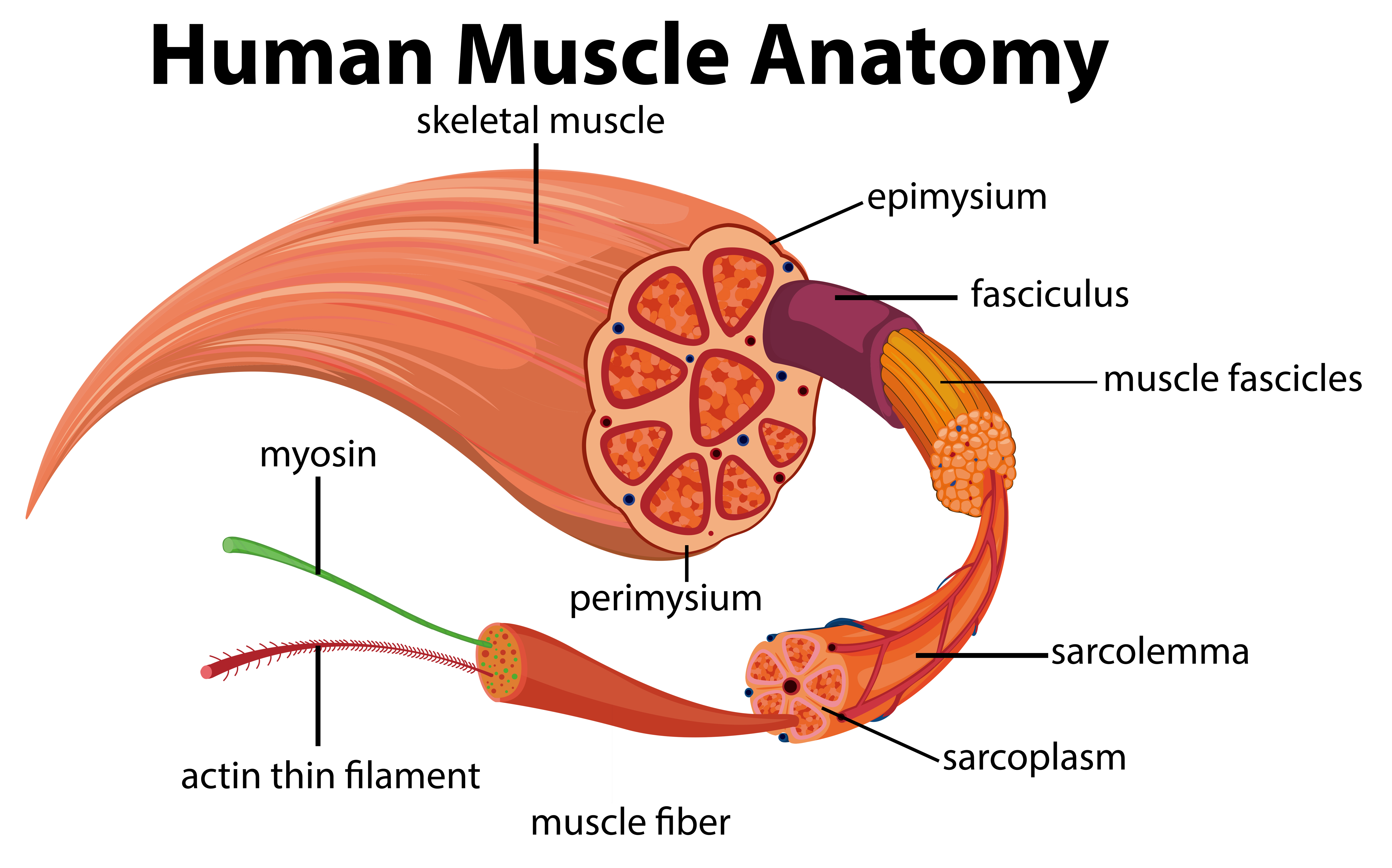
Human anatomy and physiology lab (bsb 141) module 9: The brachialis muscle lies deep to the biceps brachii, and is found more distally than the other muscles of the arm. The muscular system consists of about 700 muscle organs that are typically attached to bones across a. The muscles of the abdomen, lower back, and pelvis are separated from those of the chest by the muscular wall of the diaphragm, the critical breathing muscle. The majority of muscles in the leg are considered long muscles, in that they stretch great distances. The tendon is firmly connected to muscle fibres at one end and to components of the bone at its other end. The muscles you probably know the best are your glutes. A tendon connects the muscle to the bone. The hands enable us to perform many of our daily activities such as driving, writing and cooking. The ulnar nerve innervates the muscles of the hypothenar eminence. This is lesson 1 on the anatomy of the forearm. There are three layers of gluteal muscles on the posterior hips, just like there are three layers of muscles in the abdominal trunk. Lying exposed between the protective bones of the superiorly located ribs and the inferiorly located pelvic girdle, the muscles of this region play a critical role in protecting the.
The smaller bone that runs alongside the tibia (fibula) and the kneecap (patella) are the other bones that make the knee joint. The majority of muscles in the leg are considered long muscles, in that they stretch great distances. See more ideas about muscle anatomy, ligaments and tendons, medical anatomy. Human anatomy and physiology lab (bsb 141) module 9: Nerves and blood vessels that supply the bones and muscles of the hip.
Human Muscle Anatomy Diagram - Download Free Vectors ... from static.vecteezy.com These all work together to bear weight, allow movement and provide a stable base for us to stand and move on. The quad muscles— which form the meaty mass on the front of your thighs — are among your strongest muscle groups, and play a critical role in athletic activities. Movement occurs when our muscles pull on our bones, relocating them. The shoulder is not a single joint, but a complex arrangement of bones, ligaments, muscles, and tendons that is better called the shoulder girdle. Tendons connect the knee bones to the leg muscles that move the knee. Tendons vary in size and are somewhat elastic and attach bones to muscles. On the other hand, the insertion is where a tendon attaches that muscle to the *more* movable bone. These muscles are similar to the thenar muscles in both name and organisation.
The tendon is firmly connected to muscle fibres at one end and to components of the bone at its other end. Tendons vary in size and are somewhat elastic and attach bones to muscles. Related posts of anatomy of the foot muscles and tendons gastrocnemius muscle anatomy. The human hand is made up of the wrist, palm, and fingers and consists of 27 bones, 27 joints, 34 muscles, over 100 ligaments and tendons, and many blood vessels and nerves. Muscle anatomy forearm 12 photos of the muscle anatomy forearm forearm anatomy muscle compartments nerves, forearm extensor muscle anatomy, forearm muscle anatomy picture, forearm muscle anatomy youtube, muscle innervation forearm, human muscles, forearm anatomy muscle compartments nerves, forearm extensor muscle. This is lesson 1 on the anatomy of the forearm. Lesson on the anatomy of the forearm: Four muscles and their attached tendons make up the rotator cuff. These all work together to bear weight, allow movement and provide a stable base for us to stand and move on. The foot consists of thirty three bones, twenty six joints and over a hundred muscles, ligaments and tendons. The primary function of the shoulder girdle is to give strength and range of motion to the arm. Tendons are the connective tissues that transmit the mechanical force of muscle contraction to the bones; Foot and ankle anatomy is quite complex.

Posting Komentar
0 Komentar